
TL;DR
Medical image analysis software development means building software that ingests DICOM images and uses algorithms or AI to segment, detect and quantify findings that support diagnosis. In 2026 it combines DICOM/PACS interoperability, FHIR-based EHR integration and — for anything that aids diagnosis — SaMD compliance under FDA rules and the EU AI Act’s high-risk framework. Cost runs from $30k–$80k for a DICOM viewer to $150k–$500k+ for an enterprise AI platform.
What is medical image analysis software development?
Medical image analysis software development is the process of building software that ingests medical images — universally encoded in DICOM format from CT, MRI, X-ray, ultrasound, digital pathology or other modalities — and applies computational algorithms or AI models to segment anatomical structures, detect pathological findings, quantify biomarkers or classify findings in ways that support clinical diagnosis or treatment planning. This discipline sits at the intersection of custom healthcare software development, computer vision engineering and regulatory compliance, and it demands all three in equal measure.
The critical distinction is between imaging software (capture, store, route and display — PACS, viewers) and image analysis software (interpret, detect, measure). The first category is infrastructure; the second is where clinical AI value — and regulatory obligation — actually lives. A radiology viewer that lets a clinician pan and zoom CT slices is fundamentally different from software that highlights candidate nodules, scores them by malignancy probability and populates a structured report. Both handle DICOM; only the second is a SaMD.
The scale of the space in 2026 is significant: the FDA had authorized approximately 1,450–1,524 AI-enabled medical devices in total by early 2026, with radiology accounting for roughly 76% of that list — around 1,040–1,160 cleared algorithms — the single largest category by a wide margin, per the FDA’s published AI-enabled device list and reporting by theimagingwire.com. The FDA cleared approximately 30 AI devices per month as of early 2026, with approximately 68 new radiology algorithms cleared in Q1 2026 alone. If you are building in this category, understanding the full stack — DICOM standards, AI pipelines, FDA SaMD pathways and, for EU products, the EU AI Act’s high-risk rules — is the prerequisite.
The commercial market reflects the same growth curve: the global medical imaging software market was valued at approximately $8.11 billion in 2024 and is projected to reach $11.83 billion by 2029, a roughly 7.84% CAGR, per Mordor Intelligence analysis cited by darly.solutions — up from an earlier $2.41–$2.60 billion baseline reported for 2019–2020 by Grand View Research. AI-driven analysis and cloud-hosted deployment are the two fastest-growing sub-segments inside that figure, ahead of traditional on-premises PACS/VNA infrastructure spend.
Types of medical imaging & image-analysis software
Medical imaging software development spans several distinct product types with different architectures, regulatory footprints and build costs. The table below maps the landscape and answers the first-order regulatory question for each.
| Software type | Primary purpose | FDA clearance typically needed? |
|---|---|---|
| PACS (Picture Archiving & Communication System) | Store, index and distribute images across the organization | Often no (store/display only) |
| VNA (Vendor-Neutral Archive) | Long-term archive decoupled from any specific PACS or viewer | Often no (store/display only) |
| DICOM viewer | Clinical interface for radiologists and clinicians to view and navigate studies | Typically no (display only, unless integrated with AI analysis) |
| CADe tool (computer-aided detection) | Algorithmically detect and flag candidate findings for radiologist review | Yes — SaMD |
| CADx tool (computer-aided diagnosis) | Characterize or classify detected findings to aid diagnosis | Yes — SaMD |
| Quantification / radiomics | Measure organ volumes, lesion dimensions, radiomics features from images | Often yes if used for diagnosis or treatment planning |
| Digital pathology platform | Whole-slide image viewing, annotation and AI analysis for pathology | Yes if AI analysis aids diagnosis (IVDR in EU) |
PACS and VNA
A PACS is the backbone of an imaging department: it receives studies from modalities via DICOM, stores them, and serves them to viewing workstations and downstream systems. A VNA increasingly decouples long-term storage from the PACS vendor, providing a universal image repository that multiple PACS and viewers from different vendors can share. Modern deployments are cloud-hosted, exposing images via DICOMweb REST APIs rather than traditional DIMSE services only.
DICOM viewers
DICOM viewers are the clinical interface — the software clinicians use to open study series, navigate DICOM slices, apply windowing, perform basic measurements and generate reports. Viewers that add AI overlays or triage queues cross into the CADe/CADx territory and acquire the associated regulatory footprint.
AI/CADe–CADx analysis tools
CADe (computer-aided detection) software automatically flags candidate findings — pulmonary nodules, intracranial hemorrhage, vertebral fractures — within a study and presents them to the radiologist as a second-read tool. CADx software takes the next step: it characterizes or scores flagged findings to aid the diagnostic decision. Both constitute SaMD under FDA definitions and require regulatory clearance before clinical deployment in the US.
Core image-analysis techniques
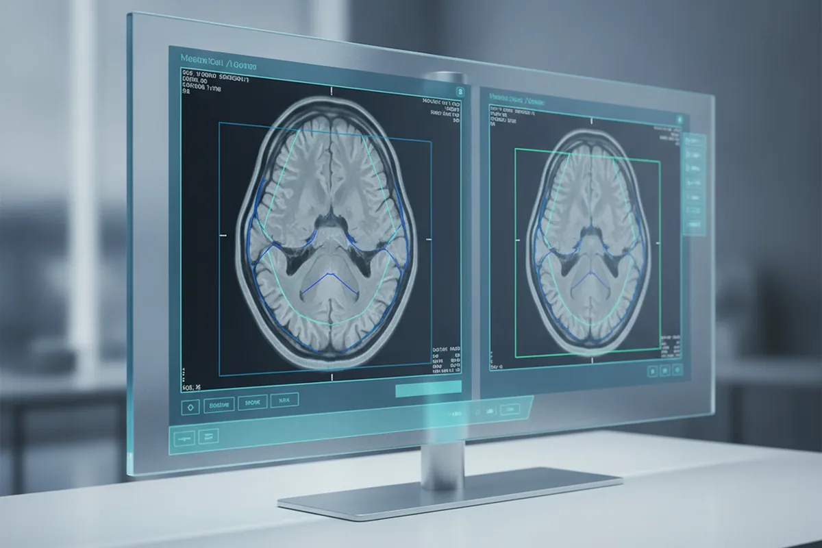
Beneath any CADe or CADx feature sits a smaller set of core image-processing techniques that recur across modalities and vendors. Which technique a given product feature requires shapes the model architecture, the annotation protocol and, often, the regulatory pathway.
| Technique | What it does | Typical use case |
|---|---|---|
| Image quality improvement | Denoising, artifact reduction, super-resolution | Make low-dose CT or motion-affected MRI diagnostically usable |
| Segmentation | Pixel-level delineation of organ or lesion boundaries | Volumetric measurement, radiotherapy and surgical planning |
| Registration / fusion | Align images from different modalities, timepoints or protocols | PET/CT fusion, longitudinal disease-progression tracking |
| Detection / localization | Flag candidate findings within a study | CADe second-read triage (nodules, hemorrhage, fractures) |
| Classification | Characterize or score a flagged finding | CADx diagnostic support (benign/malignant, severity grading) |
| Quantification | Measure size, volume or density of a structure | Tumor staging, treatment-response (RECIST) tracking |
| 3D reconstruction & 2D visualization | Build 3D models from 2D slice stacks and render clinician-facing views | Surgical planning, patient communication, MPR/volume rendering |
Modality-specific analysis: radiology, pathology, ophthalmology, cardiology
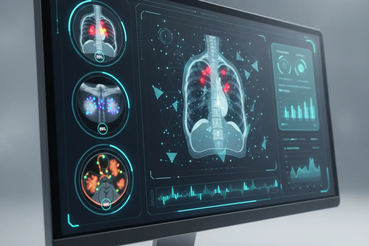
The core engineering patterns for image analysis software development are modality-agnostic — DICOM ingestion, model inference, result overlay, FHIR report output — but clinical requirements, dataset characteristics and regulatory pathways differ substantially by modality. Radiology (CT, MRI, X-ray) dominates the cleared-device landscape; digital pathology (whole-slide imaging) operates under IVDR in Europe; diabetic retinopathy screening and cardiac function analysis have their own cleared-algorithm histories and performance benchmarks.
Core standards: DICOM, DICOMweb, PACS and FHIR
Interoperability is non-negotiable for medical imaging software — every real-world deployment requires DICOM for image transport, DICOMweb for modern API access, and FHIR R4 for connecting imaging workflows to EHR clinical context.
DICOM and DICOMweb
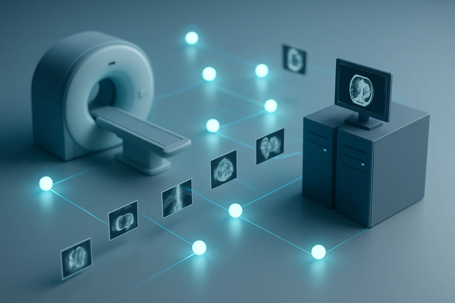
DICOM (Digital Imaging and Communications in Medicine) is the universal standard for medical image encoding, metadata and transmission. Every imaging modality — CT, MRI, X-ray, ultrasound, digital pathology scanners — produces DICOM objects. A DICOM object is not just a pixel array; it carries a structured header of patient demographics, acquisition parameters, study and series UIDs, and equipment identifiers that downstream analysis software depends on.
DICOMweb is the RESTful extension of DICOM: WADO-RS for retrieving studies and series, STOW-RS for storing them, and QIDO-RS for querying metadata — all over standard HTTPS. In 2026, committing to DICOMweb end-to-end is essential for cloud deployment, mobile access and AI inference pipelines. Legacy DIMSE services (C-STORE, C-FIND, C-MOVE) remain common in on-premises hospital infrastructure and your integration layer must handle both.
PACS versus DICOM viewer: the real difference
A PACS (Picture Archiving and Communication System) is the server-side infrastructure: it stores studies received from modalities, indexes them by patient/study/series/instance, and routes them to viewer workstations and downstream consumers (RIS, AI systems). A DICOM viewer is a client application that retrieves studies from the PACS or from a DICOMweb endpoint and renders them for clinical interpretation. The two are separate products that communicate over DICOM or DICOMweb; conflating them leads to architecture decisions that are hard to unwind later.
VNA (Vendor-Neutral Archive) sits above the PACS layer: it provides long-term, DICOM-compliant storage decoupled from any specific PACS product, allowing health systems to migrate PACS vendors without losing access to historical studies. For new enterprise deployments in 2026, a cloud-hosted VNA with DICOMweb exposure is often the recommended foundation. See our HL7 and FHIR integration guide for how imaging orders and reports connect back to the EHR layer.
FHIR R4 for EHR context
FHIR R4 (Fast Healthcare Interoperability Resources) is the standard for clinical data beyond the image itself — imaging orders (ServiceRequest), diagnostic reports (DiagnosticReport), patient demographics, allergies, prior conditions. A real medical image analysis software development project cannot treat images in isolation: the AI model’s output must flow into a FHIR DiagnosticReport that a clinician can review inside their EHR, and the imaging order context (body region, laterality, clinical indication) must flow into the analysis workflow. In 2026 this means a SMART on FHIR integration or a dedicated FHIR R4 interface alongside the DICOM layer.
How AI image analysis actually works: the model pipeline
The AI model in a medical image analysis product is only as good as the data pipeline behind it. The seven-stage pipeline below is the engineering reality of building a production-grade CADe or CADx system — each stage is a decision point that shapes both clinical performance and regulatory posture.
- Data collection and de-identification. Source DICOM studies from partner institutions, retrospective archives or prospective collections. Strip PHI from DICOM headers (de-identification per DICOM PS3.15 or HIPAA Safe Harbor) before any data leaves the clinical environment. Representation matters: collect across scanners, acquisition protocols, patient demographics and disease prevalence to avoid training bias that fails specific subgroups.
- Annotation and ground truth. Clinical annotation by qualified radiologists (or pathologists, depending on modality) is the most expensive and time-limited stage. Each finding must be labelled by clinicians with relevant subspecialty expertise; multi-reader annotation and adjudication are best practice for training data. Ground truth quality determines model ceiling — no architecture compensates for noisy labels at scale.
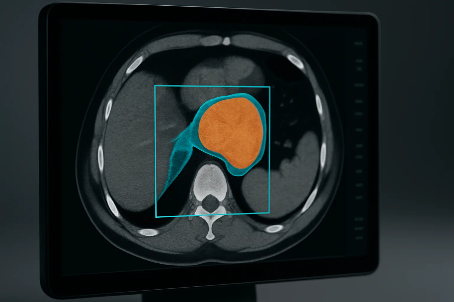
- Model selection: segmentation, detection, classification. Image segmentation (pixel-level masks for organ or lesion boundaries) is used for volumetric measurement and surgical planning. Object detection (bounding boxes around candidate findings) is the classic CADe output. Classification (yes/no, benign/malignant, severity score) is the core CADx task. Many products combine all three in a cascade: detect, then segment, then classify. Choose architecture families (U-Net derivatives for segmentation, anchor-based or anchor-free detectors for detection, CNNs or vision transformers for classification) based on the specific imaging modality and label density.
- Training and internal validation. Split your dataset into training, validation (for hyperparameter tuning) and a held-out internal test set that no model ever sees during development. Track sensitivity, specificity, AUC and clinical performance metrics (e.g., FROC for nodule detection) against the clinical benchmark for your indication. For FDA submissions, the FDA expects you to characterize performance across pre-specified subgroups: age, sex, scanner manufacturer, acquisition protocol.
- External validation. Internal validation on a held-out split from the same source data is necessary but not sufficient for regulatory submission. The FDA and EU MDR conformity assessments require performance characterization on an independent external dataset from a different institution and patient population. External validation is the step most teams underestimate and most regulators scrutinize most carefully.
- MLOps: deployment and inference. Production inference for medical imaging is GPU-bound: even a single high-resolution CT volume may contain 500+ slices and require several seconds of inference time on an optimized GPU pipeline. Deployment options are cloud (inference in HIPAA-eligible managed services, images sent via DICOMweb) or edge (GPU hardware co-located at the imaging center, no image leaving the firewall). Edge inference adds hardware procurement and update complexity but removes data-transfer latency and satisfies some health systems’ data residency requirements.
- Monitoring and drift detection. A deployed medical AI model degrades silently as scanner hardware is upgraded, acquisition protocols change or patient populations shift. Build inference monitoring that tracks prediction distribution, flags cases with low confidence for human review, and alerts when performance metrics deviate from deployment baseline. For FDA-cleared SaMD with a PCCP (Predetermined Change Control Plan), this monitoring is part of your post-market surveillance obligation.
Do you need FDA clearance? SaMD, 510(k) and PCCP
Store-and-display software alone — a PACS or a DICOM viewer that does not analyze or interpret images — typically does not require FDA clearance. Software that analyzes images or aids in diagnosis, triage or treatment decisions is classified as Software as a Medical Device (SaMD) and requires clearance before commercial distribution in the US.
SaMD classification
The FDA classifies SaMD using a risk-based framework combining the severity of the condition and the significance of the software’s role in the clinical decision. Imaging AI that provides a diagnosis or drives a treatment decision for serious or life-threatening conditions is Class III (highest risk, PMA). Imaging AI that provides information for diagnosis or treatment planning for serious conditions — the most common commercial category — is typically Class II, subject to 510(k) clearance. Class I products (inform, not diagnose, for non-serious conditions) may qualify for exemption. Classify your specific intended use honestly: the FDA scrutinizes marketing materials as much as the 510(k) submission itself. See our medical device software development guide for the full IEC 62304, ISO 13485 and ISO 14971 requirements that apply to all SaMD regardless of class.
510(k) vs. De Novo
510(k) clearance (Premarket Notification) requires demonstrating substantial equivalence to a legally marketed predicate device. For radiology AI, the predicate pool is now large enough that most commercial products can find a valid predicate; the key is matching your intended use and technological characteristics carefully to avoid a Not Substantially Equivalent (NSE) determination. De Novo review is the pathway for first-of-kind devices without a valid predicate — it results in a new product code and becomes a predicate for future 510(k)s, but adds 12–18 months and significant submission cost.
PCCP for iterating AI models post-clearance
The FDA’s PCCP (Predetermined Change Control Plan) framework, finalized in 2024 and maturing in 2026 practice, allows cleared SaMD to make defined types of AI algorithm updates — retraining on additional data within specified performance bounds, architectural changes within a defined envelope — without filing a new 510(k) for each update, provided the changes stay within the approved PCCP scope. For any medical image analysis software company building iterative AI, including a well-scoped PCCP in the original 510(k) submission is now standard practice; retrofitting it post-clearance requires a supplement.
HIPAA and PHI: building compliant imaging software
Medical images are protected health information (PHI) — they carry patient identifiers in the DICOM header and often in the pixel data itself (burned-in annotations from legacy scanners). Every component of a medical image analysis product that touches, stores, transmits or processes DICOM studies is subject to HIPAA’s Security Rule, and encryption, access control and audit logging must be engineered in from day one rather than retrofitted before launch.
The core HIPAA Security Rule requirements for imaging software: encryption of PHI at rest and in transit (AES-256, TLS 1.2+); role-based access control with minimum necessary access; immutable audit logs of all PHI access events; de-identification for any data leaving the production environment (training sets, development, testing); a signed Business Associate Agreement (BAA) with every vendor or cloud provider that processes PHI on your behalf. For a detailed checklist of every HIPAA control that medical software development requires, see our HIPAA-compliant software development checklist. For the full cost breakdown of building HIPAA compliance into a healthcare platform, see our healthcare software development cost guide.
EU AI Act & MDR: why medical imaging AI is high-risk
Imaging AI that is embedded in or constitutes a medical device regulated under the EU Medical Device Regulation (MDR) or In Vitro Diagnostic Device Regulation (IVDR) is automatically classified as high-risk under the EU AI Act — this covers virtually all commercial diagnostic imaging AI for the EU market.
The scope is broad: AI systems intended to influence clinical decisions about patients using medical devices, or which are themselves devices under MDR/IVDR, fall squarely in Annex III (high-risk AI). For imaging AI, this means any CADe, CADx or quantification tool used in a clinical diagnostic context. Most commercial imaging AI products operate at EU MDR Class IIa or higher (the conformity class is determined by the MDR rules for medical devices, not by the AI Act itself).
High-risk AI obligations under the EU AI Act include: a quality management system; comprehensive technical documentation; data governance and dataset documentation; transparency requirements (users must know they are interacting with an AI system); human oversight mechanisms (the AI output must be reviewable and overridable by a qualified clinician); accuracy, robustness and cybersecurity measures; and post-market monitoring. The good news for MDR-regulated products: these obligations are assessed jointly with MDR conformity by the Notified Body in a single assessment, not as a separate second track. This means if your MDR technical file and post-market surveillance plan are solid, the AI Act obligations are largely covered by the same body of evidence.
As of 2026, the applicable compliance deadlines have shifted from earlier projections: the EU AI Act’s high-risk duties for standalone high-risk AI systems are expected by 2 December 2027; for AI systems embedded in regulated products (including MDR/IVDR medical devices), the deadline is 2 August 2028, per medtecheurope.org (2026-05), reedsmith.com and dqsglobal.com commentary. Development teams should nonetheless design for these obligations now — retrofitting data governance, human oversight mechanisms and technical documentation into a shipping product is substantially more expensive than building for them during initial development.
Architecture & technology stack (2026)
A modern medical image analysis stack in 2026 is cloud-native, standards-first and GPU-backed for inference. The table below maps each layer to the typical technology approach; specific vendor products vary by client context, budget and data residency requirements.
| Layer | Typical approach / tools (2026) |
|---|---|
| Image ingestion | DICOM DIMSE (C-STORE from scanners/PACS) + DICOMweb STOW-RS for cloud push; de-identification on ingest |
| Storage (PACS/VNA) | Cloud-hosted DICOMweb-compliant VNA (HIPAA-eligible object storage with DICOM indexing); legacy on-premises PACS bridged via DICOM gateway |
| DICOM viewer | Web-based viewer (OHIF/Cornerstone.js pattern): DICOMweb WADO-RS for study retrieval, WebGL rendering; client-side zero-footprint for hospital firewall environments |
| AI model training | PyTorch with MONAI-class medical imaging libraries; segmentation (U-Net variants), detection (nnDetection or DETR-class), classification (ResNet/ViT); MLflow or equivalent for experiment tracking |
| AI inference | Cloud: GPU-accelerated inference endpoint (NVIDIA T4/A10G class) in HIPAA-eligible VPC; Edge: NVIDIA Jetson or dedicated workstation GPU co-located at imaging center; ONNX or TorchScript for deployment format |
| Interoperability (FHIR/HL7) | FHIR R4 DiagnosticReport for AI findings; SMART on FHIR for EHR context; HL7 v2 ORU messages for legacy radiology information systems |
| Annotation & data ops | DICOM-SR or DICOM SEG for structured annotations; label management platform; data versioning; de-identification pipeline |
| Security & compliance | AES-256 at rest, TLS 1.3 in transit; RBAC; immutable audit logs; HIPAA-eligible cloud region and signed BAAs; IEC 62304 software lifecycle documentation |
Development process: from discovery to clinical validation
A compliant medical imaging software development engagement follows a predictable sequence — the stages teams rush (regulatory scoping and data strategy) are where the clinical and commercial outcome is decided, not the model architecture.
- Discovery and regulatory strategy (3–5 weeks): define the clinical intended use precisely, determine whether the product is SaMD, identify the FDA pathway (510(k) or De Novo) and the EU MDR class, and map data requirements for the indication. This stage should produce a written regulatory strategy before a line of model code is written.
- Data strategy and collection (ongoing, 4–12+ weeks): identify data partners, negotiate data use agreements (DUAs), run de-identification validation, and design the annotation protocol. For rare conditions, multi-site collection and a federated learning architecture may be needed to reach clinically meaningful dataset size.
- Prototype viewer and integration (4–8 weeks): build the DICOM ingestion pipeline, DICOMweb integration to the target PACS or VNA, and an initial viewer overlay for AI outputs. This validates the infrastructure before model development consumes most of the timeline.
- Model development and internal validation (8–20 weeks): iterative training and evaluation against the held-out internal test set, with performance tracked against clinically meaningful benchmarks for the indication. Architectural and hyperparameter decisions are locked for the validation dataset before it is scored.
- External validation and clinical study (8–16 weeks): independent dataset, often from a partner institution; reader study design (standalone vs. with-AI comparison); IRB approval where required. External validation data is what populates the clinical performance section of a 510(k) submission.
- Regulatory submission (510(k): typically 3–6 months FDA review; EU MDR: 6–18 months Notified Body): submit the 510(k) with design controls documentation (IEC 62304 lifecycle), software description, validation protocol and report, clinical study data, and PCCP if included. Respond to additional information (AI) requests promptly.
- Deployment, monitoring and post-market surveillance (ongoing): cloud or edge deployment; MLOps monitoring for drift; adverse event reporting per FDA MDR (Medical Device Reporting) rules; post-market clinical follow-up (PMCF) per EU MDR 2017/745.
How much does medical image analysis software cost in 2026?
Medical image analysis software development cost in 2026 runs from roughly $30,000–$80,000 for a basic DICOM viewer with PACS integration, to $150,000–$500,000+ for a full enterprise AI imaging platform with custom model pipeline, EHR integration and regulatory compliance architecture. These are 2026 market estimates from appzoro.com and corpsoft.io; the final number is driven by regulatory class, integration complexity and dataset size more than visible feature count.
| Scope | Typical range (2026) | Primary cost drivers |
|---|---|---|
| DICOM viewer + PACS integration (no AI analysis) | $30k–$80k | DICOMweb integration complexity, number of modalities, HIPAA controls |
| Single-modality AI detection or classification feature (CADe/CADx) | $80k–$200k | Dataset annotation cost, model validation, 510(k) preparation |
| Multi-modality AI platform (FDA-cleared, EHR integration, cloud) | $150k–$500k+ | Multi-site data, external validation, regulatory submission fees, FHIR integration, MLOps infrastructure |
| Enterprise deployment with EU MDR conformity + FDA clearance | $300k–$800k+ | Dual regulatory pathways, Notified Body fees, clinical study, post-market surveillance infrastructure |
Regulatory submission fees are a separate line item: a standard 510(k) FDA user fee in 2026 is approximately $24,000–$26,000 for most applicants. Notified Body assessment costs for EU MDR Class IIa certification typically run $40,000–$100,000+ depending on the body and the scope of the technical file. Neither is included in the development cost estimates above. For a broader view of healthcare software development cost, see our healthcare software development cost guide.
Key challenges in medical image analysis software development
The core technical and operational challenges that account for most project overruns and product failures in this space:
- Data scarcity and class imbalance. Rare pathologies by definition have few labelled cases in any single institution’s archive. A model trained on a skewed dataset produces unreliable sensitivity for minority classes; correcting this in post-market performance data is far more expensive than designing for it from the outset with multi-site collection and federated learning.
- Annotation cost and inter-reader variability. Expert radiologist time is the scarcest and most expensive input. Inter-reader disagreement on boundary delineation and finding characterization introduces irreducible label noise. Adjudication protocols and consensus-labelling workflows must be built into the data pipeline, not treated as a one-off effort.
- Generalization and external validation failure. Models that achieve 0.95 AUC on internal test data routinely degrade to 0.78–0.85 on external datasets from different scanners, protocols and populations. Designing for robustness — multi-site training, protocol augmentation, scanner-agnostic normalization — is engineering work, not luck.
- PACS and EHR integration friction. Every hospital PACS installation is different: custom DICOM configurations, non-standard DICOM tags, legacy DIMSE-only interfaces. EHR sandboxes are slow to provision and production credential timelines can extend the deployment phase by months. Build integration time into every milestone plan.
- Regulatory lead time. A 510(k) with additional information (AI) requests can take 12+ months from submission to clearance. EU MDR Notified Body timelines have stretched to 18–24 months for complex products. Teams that start regulatory preparation after model development is complete routinely miss commercial launch windows by a year or more.
- Clinician trust and workflow fit. A technically validated AI tool that interrupts the radiologist’s reading workflow, produces too many false positives or adds screen-switching friction achieves low adoption regardless of clearance status. Co-design with radiologists, embed the AI output in the existing workflow rather than a separate screen, and instrument adoption metrics from day one of deployment.
- Post-market drift and regulatory obligation. A cleared AI device has ongoing obligations: FDA Medical Device Reporting (MDR) for adverse events, periodic performance reporting, and — for PCCP holders — defined monitoring thresholds that trigger either a PCCP-bounded update or a new submission. Build the monitoring infrastructure as part of the initial platform, not as a Phase 2 backlog item.
FAQ
What is medical image analysis software development?
Medical image analysis software development means building software that ingests DICOM medical images from CT, MRI, X-ray, ultrasound or pathology scanners and applies algorithms or AI to segment, detect, quantify or classify findings that support clinical diagnosis or treatment planning. It combines DICOM/PACS interoperability, FHIR-based EHR integration and — for anything that aids diagnosis — SaMD compliance under FDA rules and the EU AI Act’s high-risk framework.
Does medical imaging software need FDA clearance?
Store-and-display software (a PACS or DICOM viewer that does not analyze or interpret images) often does not require clearance. Software that analyzes images or aids in diagnosis, triage or treatment decisions is classified as SaMD and requires clearance — most commonly via 510(k) for Class II products, or De Novo for novel first-of-kind devices. HIPAA and IEC 62304 apply regardless of clearance status.
How much does it cost to build medical image analysis software in 2026?
A DICOM viewer with PACS integration runs $30k–$80k. A mid-tier product with a single AI detection or classification feature runs $80k–$200k. A full enterprise imaging platform with custom AI pipeline, EHR integration, cloud infrastructure and FDA/MDR compliance runs $150k–$500k+. Regulatory submission fees (FDA 510(k): ~$24k–$26k; EU MDR Notified Body: $40k–$100k+) are additional. Cost is driven by regulatory class, dataset annotation, integration complexity and clinical validation scope.
What is the difference between PACS and a DICOM viewer?
A PACS is server-side infrastructure that stores, indexes and distributes medical images across an organization. A DICOM viewer is a client application that retrieves studies from the PACS or a DICOMweb endpoint and renders them for clinical interpretation. A Vendor-Neutral Archive (VNA) decouples long-term image storage from the PACS vendor, allowing multiple viewers and PACS products to share a single repository.
Is medical imaging AI high-risk under the EU AI Act?
Yes — virtually all commercial diagnostic imaging AI for the EU market. AI embedded in or constituting a medical device regulated under EU MDR or IVDR is automatically classified as high-risk under the EU AI Act. High-risk obligations (data governance, transparency, human oversight, accuracy documentation) are assessed jointly with MDR/IVDR conformity by a Notified Body. Applicable deadlines have shifted to 2 December 2027 (standalone high-risk AI) and 2 August 2028 (AI embedded in regulated products), per medtecheurope.org (2026-05).
What tech stack is used for medical image analysis software?
A modern 2026 stack: DICOMweb (WADO-RS/STOW-RS/QIDO-RS) for image transport; a DICOM-compliant VNA or cloud PACS for storage; an OHIF/Cornerstone.js-style web viewer for the clinical interface; PyTorch with MONAI-class libraries for model training; GPU-accelerated inference (cloud or edge); FHIR R4 for orders and reports to/from the EHR; and HIPAA-compliant encryption, RBAC and audit logging throughout.
Last updated 27 September 2026. Cost ranges are 2026 market estimates from appzoro.com and corpsoft.io; FDA device counts per radiologybusiness.com, auntminnie.com and theimagingwire.com (2026-03); market size per Mordor Intelligence via darly.solutions and Grand View Research; EU AI Act deadline guidance per medtecheurope.org (2026-05), reedsmith.com and dqsglobal.com. Not legal or regulatory advice — consult a qualified regulatory affairs specialist for your specific product and intended use.